Petar Raychev
Sofia University “St. Kliment Ohridski”
https://doi.org/10.53656/str2025-2s-9-hex
Abstract. In the higher education system, modern physiology courses play a crucial role in preparing future specialists in human and veterinary medicine, sports, and various areas of medical and biological sciences. Physiology is an experimental science and electrophysiological methods play a leading role in its development and application in practice. Electrocardiography is the most widely used electrophysiological method in clinical and sports physiology and medicine. For this reason, it is traditionally and invariably included in the teaching content of almost all modern physiology courses. From a didactic point of view, vector analysis in electrocardiography is one of the most problematic areas in physiology education. The conceptual apparatus and didactic approaches in the presentation of the hexaxial system and its application in the analysis and interpretation of electrocardiographic recordings are particularly important for the quality of the learning process. Based on a content analysis of contemporary educational literature, the study outlines important problematic points related to the applied conceptual apparatus in the didactic process of this topic. A didactic model aims to overcome the identified problems and increase the effectiveness of the physiology teaching process in its section on vector analysis in electrocardiography.
Keywords: physiology training; electrocardiography; human physiology; qualitative inspection method; hexaxial system; ECG vector analysis
Introduction
Physiology is one of the most challenging disciplines in the medical education system, and it has become the cause of dropout for many students enrolled in medicine (Michael 2007; Slominski et al. 2019). Many general and specific methodological problems in the didactics of physiology, related to the construction of systems of interrelated and logically consistent concepts, contribute significantly to this.
At the present stage of its development, animal and human physiology is an integrative dynamic science with a clear experimental character (Raychev 2024). This requires in the teaching and learning of physiology, considerable attention be paid to the methods underlying its development and practical application.
In a broader sense, electrocardiography is an electrophysiological method in which the total bioelectric activity of the heart is recorded, analyzed, and interpreted. It is the most widely used electrophysiological method in clinical practice, sports physiology, etc. For this reason, electrocardiography as a topic is present in the physiology curriculum, not only for future physicians (Barret et al. 2019; Boron & Boulpaep 2017; Brandes et al. 2019; Conti 2020; Costanzo 2018; Fox 2019; Hall & Hall 2021; Kibble 2020; Khurana & Khurana 2015; Koeppen & Stanton 2024; Pal 2017; Rhoades & Bell 2018; Sembulingam & Sembulingam 2012; Sherwood & Ward 2019; Silverthorn 2019; Stanfield 2017; Widmaier et al. 2023; Alipov 2013; Vitanova & Garchev 2020; Smirnov 2016), but also for sports professionals (Draper et al. 2024; Kenney et al. 2012; Kraemer et al. 2012; Plowman & Smith 2014) and a range of other fields. Introducing the basic principles of electrocardiography to future professionals is therefore an important task of physiology courses. In this context, it is striking that the physiology teaching literature contains a wide variety of approaches to the scope, structuring, and didactic presentation of this topic. These range from very superficial and fragmentary references (Costanzo 2018; Fox 2019; Widmaier et al. 2023) to attempts at a relatively comprehensive and coherent presentation of the fundamentals of electrocardiographic method (Boron & Boulpaep 2017; Conti 2020; Hall & Hall 2021; Khurana & Khurana 2015; Pal 2017; Rhoades & Bell 2018).
A key challenge for students in the Electrocardiography topic is the introduction to vector analysis. For them, it is a kind of ‘high-flying’ form that has important practical implications for mastering the analysis and interpretation of electrocardiograms. However, in the current educational literature on physiology, vector analysis is usually ignored (Costanzo 2018; Fox 2019; Sherwood & Ward 2019; Silverthorn 2019; Stanfield 2017; Widmaier et al. 2023; Vitanova & Garchev 2020) or presented sparsely and incompletely (Barrett et al. 2019; Conti, 2020; Koeppen & Stanton 2024; Sembulingam & Sembulingam 2012). Few authors have attempted a more comprehensive and logically consistent coverage of the topic (Boron & Boulpaep 2017; Hall & Hall 2021; Pal 2017; Rhoades & Bell 2018; Alipov 2012).
A major problem in mastering vector analysis is the lack of a logically coherent, unified and widely accepted conceptual framework that can be effectively applied in the didactic process. This leads to difficulties in structuring and organising the teaching content. A leading example in this respect is the notion of a ‘hexaxial system’. Mastering the basics of vector analysis requires a comprehensive and logically coherent concept of the hexaxial system to be applied in the learning process.
Methods
The model is based on a scientific-didactic and structural-logical analysis of the content of modern physiology courses presented in current educational literature. The organisational centers of the analysis are the scientific reliability of the presented information, its correspondence with the educational needs of the audience, and the didactic notion of the subject as a system of concepts structured according to a certain model.
The model is designed taking into account mostly the educational needs of medical students in their human physiology course.
The relevance of the analyses and conclusions is determined by the literature sample, which mainly includes publications after 2010. The fact that the literature sample mostly includes sources that have gone through at least two editions contributes to a higher degree of representativeness, validity, and quality of the educational literature used.
Didactic model of formation of the concept of “hexaxial system” and practical skills for its application
Effective and lasting mastery of vector analysis and subsequent formation of practical skills for its application strongly depend on the system of concepts formed in the learning process and the degree of their integration into a common theoretical framework. Key concepts are “electrode”, “conduction”, “electrical axis of the heart”, “mean electric axis of the heart”, “Einthoven’s triangle”, “hexaxial system”, etc.
Hexaxial system. Basic concepts in modern teaching literature
Electrode. The term electrode has a pivotal importance in electrophysiology, but it is rarely defined as a concept in physiology courses. This is also true of literature dealing specifically with electrophysiology. In the context of electrocardiography, it is interesting to note that an electrode is defined by some authors (Pal 2017, p. 760) as an electrical conductor placed on the heart or body surface and used in combination with another such conductor to record potential differences in the heart.
In electrophysiology, particularly in electrocardiography, the terms active (or recording) electrode and indifferent electrode are widely used (Barrett et al. 2019; Khurana & Khurana, 2015; Pal 2017). However, they are almost always present in the physiology education literature only as terms and not as clearly defined concepts. Pal (2017) defines indifferent electrodes as a reference input. However, this confuses different entities and concepts (electrodes and parts of electrocardiographic machines).
For bipolar leads, the terms positive and negative electrode are very commonly used, which is highly misleading for trainees (Pal 2017, p. 760; Sembulingam & Sembulingam 2012, p. 552), who perceive them as cathode and anode.
Lead. The concept of lead is pivotal in electrophysiology. However, despite using the term, many authors prefer not to define it as a concept (Costanzo 2018; Kibble 2020; Barrett et al. 2019; Vitanova & Garchev 2020). In physiology textbooks that do define lead, there are inconsistencies and no clear distinction between electrodes and leads as concepts.
Some authors divide leads into monopolar and bipolar according to the number of active electrodes in them: one or two (Khurana & Khurana 2015). In bipolar leads, one electrode is connected to the positive terminal and the other to the negative terminal of the electrocardiograph, implying that the poles of the lead are ‘determined’ by the machine recording the heart’s bioelectric activity.
According to some authors (Sembulingam & Sembulingam 2012), the lead is one electrode, which contradicts the statement on another page of the same textbook that in bipolar leads there is a coupling of two terminals, and when considering unipolar leads, two electrodes are mentioned, one active and one indifferent. For other authors (Boron & Boulpaep 2017; Hall & Hall 2021; Khurana & Khurana 2015; Pal 2017; Silverthorn 2019), the lead is a pair of electrodes attached to the body. According to Stanfield (2017, p. 404), a lead is a pair of electrodes denoted by Roman numerals (i.e., only Einthoven leads). A logical consequence of this understanding of leads is the use of the term limb leads (Rhoads & Bell 2018; Pal 2017; Sherwood & Ward 2019). According to Fox (2019), leads are the active electrodes. Such a thesis is also expressed in some textbooks on electrocardiography (Bayes et al. 2022). The paradoxical thesis is that there are six bipolar leads and that, for example, the electrode on the left arm (L) is simultaneously two leads. The main problem with definitions that confuse or do not clearly distinguish electrodes from leads is that they are incompatible with classical concepts of leads and Einthoven’s triangle. For unipolar leads, they create a contradiction between the number of electrodes (only 3 for frontal and 9 for precordial entries) and the number of leads (6 frontal and 6 precordial). Thus, the introduction of the concept of a monopolar lead creates a logical and factual contradiction, effectively redefining the concept of a lead within the same training text (Pal 2017; Fox 2019).
According to Vander’s textbook (Wildmaier et al. 2013, p. 377), leads are multiple combinations of recording sites located on the limbs and thorax. The problem here is that no distinction is made between an electrode fixation scheme (standard) and a lead. It is an interesting idea to think of a lead as a system of electrodes connected to produce a single electrocardiogram recording (Sherwood & Ward 2019, p. 358).
Lead pole. The term ‘lead pole’ (positive and negative) is used in some texts to describe bipolar leads (Rhoades & Bell, 2018; Hall & Hall 2021). For unipolar leads, the negative pole is replaced by a point in the center of the chest with zero potential. At the same time, terms such as the positive and negative terminals of the electrocardiograph are also used when describing bipolar leads and Goldberger leads (Hall & Hall 2021 pp. 139-140). In the same textbook, when discussing vectorcardiograms, it is pointed out that the zero-reference point is the negative terminal of all subsequent vectors. This last statement can be adapted to Goldberger’s unipolar leads and thus form a wider meaning and application of this concept in the didactic process.
Lead axis. The term ‘lead axis’ is used in conjunction with the terms ‘Einthoven triangle’ and ‘hexaxial system’. The main problem lies in the fact that it is used precisely as a term and not as a concept with clearly defined content and scope (Barrett et al. 2019; Pal 2017; Silverthorn 2019; Vitanova & Garchev 2020). According to Boron & Boulpaep (2017), each lead is an axis lying in a specific plane in which the electrical activity of the heart is projected, i.e. axis and lead are identical. The lead axis is defined by Kibble (2020) as a unique property or component of the lead. According to Guyton’s textbook (Hall & Hall 2021), the lead axis is the direction from the negative to the positive electrode of the lead, which only applies to bipolar leads. Here (p. 143), a meaningful discrepancy exists between a term (‘axis’) and a concept (‘direction’).
Einthoven’s triangle. In electrocardiography, the idea of Einthoven’s triangle is fundamental. As many physiology textbooks only partially or not at all cover vector analysis of the ECG, both Einthoven’s triangle and the hexaxial system are often omitted from the teaching content (Costanzo 2018; Fox 2019). Typically, the Einthoven triangle is defined as an equilateral triangle lying in the frontal plane of the body, with its sides corresponding to the three bipolar lead axis and the heart at its center (Barrett et al. 2019; Khurana & Khurana 2015). This definition is consistent with the original publication by Einthoven (1912). The base of the triangle lies between the two arms and its apex points towards the groin (Koeppen & Stanton 2024), with two active electrodes placed at two corners of the triangle (Khurana & Khurana 2015). Guyton’s textbook (Hall & Hall 2021 p. 139) makes the important point that the vertices of the triangle are like points where the limbs connect to the fluids surrounding the heart.
Another textbook (Rhoades & Bell 2018) draws attention to Einthoven’s triangle as a convention. However, one problem is the representation of the vertices of the triangle as electrode attachment points rather than as pole positions of the bipolar leads. Ganong’s textbook (Barrett et al. 2019) also proposes the vertices of the triangle as electrode attachment sites.
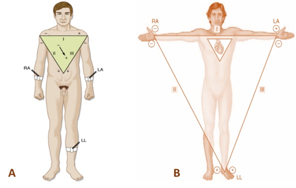
Figure 1. Types of Einthoven's triangle illustrations frequently used in physiology textbooks. А. Accurate representation of the position of the triangle according to the original model. В. Incorrect and logically contradictory representation of Einthoven's triangle. The association of the vertices of the triangle with the positions of the electrodes is misleading for the learner. The superimposition of two triangles exacerbates the problem.
The graphical representation of Einthoven’s triangle is important in the didactic process. In line with the classical model, illustrations such as the one shown in Figure 1A are both logically and didactically valid (Boron & Boulpaep 2017; Hall & Hall 2021; Koeppen & Stanton 2024; Khurana & Khurana 2015). On the other hand, it is scientifically incorrect and didactically problematic to present Einthoven’s triangle in illustrations similar to Figure 1C (Stanfield 2017; Sherwood & Ward 2019).
Hexaxial system of frontal leads. The hexaxial system is presented whenever the basics of vector analysis are explained (Hall & Hall 2021; Khurana & Khurana 2015; Rhoads & Bell 2018; Boron & Boulpaem 2017, etc.). In the didactic process, it is usually ‘derived’ based on Einthoven’s triangle by combining the axes of the standard frontal leads so that they intersect at a single point (Figure 2A). An interesting idea in the didactic process is to use the analogy between the hexaxial system and the face of a clock (Khurana & Khurana 2015).
Electric axis of the heart. Many modern physiology textbooks do not define the electric axis of the heart. Some authors define the electric axis of the heart as “the line connecting the points with the greatest potential difference at a given moment…” (Logofetov 1997). In Guyton’s classic textbook (Hall & Hall 2021), the electric axis of the heart is called the “instantaneous sum vector”. Similar definitions are given by other authors (Barrett et al. 2019; Vitanova & Garchev 2020). According to some authors (Pal 2017; Alipov 2013), this vector is derived from the instantaneous summation of all dipoles in the heart. There are textbooks (Khurana & Khurana 2015) in which the electric axis of the heart is represented as an instantaneous vector that reflects the magnitude and direction of the electrical potential during the cardiac cycle. The thesis of the electrical axis of the heart as a sum vector is also found in the Bulgarian educational literature (Vitanova & Garchev 2020).
Mean electric axis of the heart. The mean electric axis of the heart is defined as the average value of the electric axis of the heart during ventricular depolarisation (Alipov 2013), which creates a discrepancy between the term used and the content of the concept, referring only to a short period of the cardiac cycle. Some textbooks use the mean electric axis of the heart without defining the electric axis of the heart (Koeppen & Stanton 2024).
In the text by Rhoades (Rhoades & Bell 2018), two important points are made. First, the mean electric axis of the heart is a vector, and second, it is the average of the electric axis of the heart throughout the ventricular depolarisation (QRS complex). The authors use the term “mean QRS electric axis”. Other authors use similar terminology (Pal 2017; Barrett et al. 2019; Hall & Hall 2021). Guyton’s textbook (Hall & Hall 2021) also uses the term “mean electric ventricular axis”. In the same textbook, the use of the term “mean T-wave axis” is interesting, but is not accompanied by any further explanation. The term “wave axis in the frontal plane” can be highlighted as a significant step forward in didactic practice, which is also not framed as a concept (Boron & Boulpaep 2017).
A key attribute of the mean electric axis of the heart, with important diagnostic significance, is the value of the angle it makes with I+. In this respect, from a didactic point of view, the inclusion in the teaching content (Boron & Boulpaep 2017, p. 498; Pal 2017, p. 770) of the qualitative inspection method for estimating the angle of the central electrical axis can be regarded as productive.
Description and algorithm of the didactic model
The model aims to present the basic concepts underlying the idea of the hexaxial system in electrocardiography and to form skills for its application in the analysis of electrocardiographic recordings. It can be implemented in the linear algorithm described below.
Step 1. Introducing the concept of “lead”. It can be defined as a set of two or more electrodes connected in a specific way to the electrocardiographic machine and used for a single registration of the total bioelectrical activity of the heart. It is assumed here that concepts such as electrode, electrode types (as specific electrophysiological concepts!), electrocardiograph, and electrocardiogram have already been formed.
Step 2. Defining the concept of lead pole. In this step, the following important points should be emphasised. Each standard lead is characterised by two imaginary points associated with a particular region of the body, called poles. One pole is called positive and is associated with the positive input of the electrocardiograph. The other is termed negative or zero (indifferent) and is connected to the negative input of the electrocardiograph. Pole designations are defined by convention of the scientific and expert communities and relate to how the bioelectrical activity of the heart is represented. It is important to emphasise to trainees that these symbols are not related to the nature of electrical charges or to the concepts of cathode and anode, which are important in the methods of electrical stimulation of different tissues and organs. It has to be emphasized also, that the poles of the lead are not identical to the attachment points of the electrodes and should not be confused with them.
Step 3. Defining the concept of a lead axis. A lead axis can be defined as a mental straight line connecting the poles of a lead.
Step 4. Defining the term “bipolar lead”. A bipolar lead can be thought of as two active electrodes connected to the lead’s poles (+ or -).
Step 5. Defining the term “monopolar lead”. A monopolar lead can be considered a lead with one active electrode connected to the positive pole of its axis (i.e., the positive input of the device). Two or more electrodes are attached to the negative pole.
Step 6. Presentation of the main types of ECG leads according to the plane of their axes (frontal and precordial).
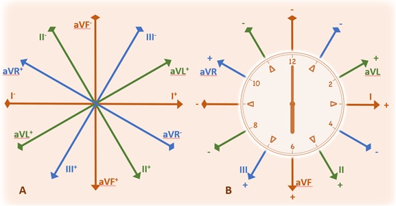
Figure 2. Illustrations for development of the concept of "hexaxial system" in the didactic process of physiology.. А. The hexaxial system is represented with emphasis on its orthogonal axes of leads. В. Demonstration of the analogy of the hexaxial system with the clock face
Step 7. Familiarization with Einthoven’s triangle. An important point in the didactic process here is to avoid significant deviation (in description and illustration) from Einthoven’s classic triangular model (Figure 1A). It is especially important to position the triangle correctly with the heart and body.
Step 8. Introducing the unipolar Goldberger leads. Demonstrate the position of their axes and poles in the Einthoven triangle in combination with those of the bipolar leads. Introduce the principles of electrode coupling in these leads.
Step 9. Deriving the hexagonal system of frontal leads from the Einthoven triangle. In this step, the derivation of the hexaxial system and the Cabrera circle from the Einthoven triangle is demonstrated with appropriate illustrations.
Step 10. Demonstration of the analogy between the hexaxial system and the face of a clock. It is appropriate to choose an illustration with a dial analogous to that shown in Figure 2B.
Step 11. Familiarisation with the rules for expressing angles in the hexaxial system to the positive semi-axis of the first lead (I+).
Step 12. Pointing out and listing the pairs of lines that are orthogonal (perpendicular to each other) in the hexaxial system. Emphasise that in these pairs one lead axis is bipolar and the other is unipolar.
The steps up to this point provide a coherent understanding of the hexaxial system. It will serve as a basis for applying vector analysis to electrocardiographic recordings.
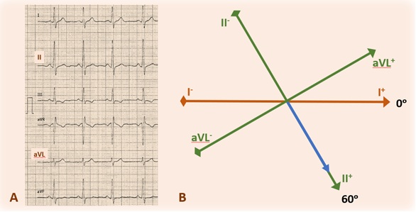
Figure 3. A typical illustration that can be used to build skills in determining the mean electric axis of the heart for the period of ventricular depolarization. The relationship between the electrocardiogram (A) and the hexaxial system (B) is shown. It can be seen that the axis is perpendicular to aVL. Accordingly, the projection vector and amplitude of the QRS complex is greatest in lead II. The slope is approximately 60ᵒ
Step 13. Defining the concept of the „electric axis of the heart“. It is important to introduce it as a vectorial quantity, i.e. a quantity with magnitude and direction. One of its essential characteristics is that it is a quantity obtained by summing a series of vectors located on the de/repolarization wavefront.
Step 14. Definition of the concept of the mean electric axis of the heart for the main components of the electrocardiogram (P wave, T wave, and QRS complex). Depending on the time available and the organization of the training, students can be introduced to the procedure for determining the angle of the ventricular depolarization period from the QRS complexes of two bipolar leads (e.g., I and II). A diagram adapted to the learning process can be used for this purpose.
Step 15. Formation of the skill to determine the angle of the mean electric axis of the heart for the period of ventricular depolarization. The following algorithm can be used to find the mean electric axis of the heart for the period of ventricular depolarization:
A. On the electrocardiogram, identify the lead whose axis is isoelectric (perpendicular) or nearly isoelectric to the mean electric axis. This is evident from the fact that the amplitude of the QRS complex is either zero or of minimal value compared to the amplitudes in all the other frontal leads (aVL in Figure 3A).
B. Look at the lead with an axis orthogonal to the lead axis identified in the previous step (II in Figure 3A). The recording has the maximum amplitude of the QRS complex in absolute value relative to the other leads in the same polarity group (Einthoven or Goldberger leads). The axis of this lead appears to be parallel or nearly parallel to the mean electric axis of the heart.
C. Determine the orientation of the projection vector on the axis of the parallel lead (+ or -) according to whether the amplitude of the QRS complex is positive or negative. In the case of Figure 3A, it is II+.
In the first three steps, the ability to estimate the orientation of the mid-electrical axis is formed by applying the qualitative inspection method. Extending this method, in the next steps, the ability for a more accurate estimate of the angle of the heart’s mean electric axis is formed.
D. Assess the amplitude of the QRS complex in the isoelectric lead as positive, zero, or negative.
E. If the amplitude value is zero, the angle of the mean electric axis coincides with the angle (90ᵒ) of the half-axis of the orthogonal lead on which its projection vector lies (as in Figure 3). If the amplitude value is not zero, 5ᵒ (if the difference is barely noticeable) or 10ᵒ (if the difference is more pronounced) is added to or subtracted from the angle of the corresponding half-axis of the orthogonal lead with I+.
F. The estimated value of the angle of the mean electric axis is compared with the automatically calculated value on the electrocardiogram. For the given example in Figure 3A, it is 59ᵒ.
Step 16. Forming the ability to interpret the angle of the mean electric axis of the heart for the period of ventricular depolarization. The trainees are familiarized with the basic normal and pathological angle intervals. It is convenient to use an appropriately selected illustration.
Diagnosis of the learning content acquisition
Three types of tasks can be used to diagnose the effectiveness of the didactic process within the described model and the level of learning of the subject content.
The first type of tasks checks the mastery of the six-axis system. They check the students’ knowledge of the angles of the half-axes of the lines and their polarity (+ or -). An example of this type of problems is the question „What is the angle of II+?
In the second type of problems, the candidate is asked to find the angle of the mean electric axis of the heart from a given electrocardiogram and to interpret its value. The candidate must be able to explain how they determined the angle of the axis.
In the third category of problems, the student must use the value of the angle of the mean electric axis of the heart to determine in which lead the amplitude of the QRS complex has a maximum and a minimum value. An example of this type of problems is: „If the angle of the mean electric axis of the heart is 60⁰, in which lead will the QRS complex have maximum and minimum amplitude?
Discussion and recommendations
The main focus of the didactic model described above is the formulation of a system of interrelated concepts, based on which the concept of the hexaxial system will be developed. A key element in this is the development of a concept of lead poles. This is a theoretical construct necessary to form an overall idea of the lead concept, including the electrocardiograph inputs, the position and number of electrodes, and the construction of different types of leads. This overcomes the problems in the didactic process associated with the practice of defining leads based on electrodes, most commonly on the principle of one lead-two electrodes (Boron & Boulpaep 2017; Hall & Hall 2022; Khurana & Khurana 2015; Pal 2017; Silverthorn 2019; Stanfield 2017). The analogy between the hexaxial system and the clock face as a didactic approach aims to increase the effectiveness of the learning process. Using an expanded form of the qualitative inspection method allows students to acquire the skills to more accurately assess the angle of the mena electric axis of the heart.
The diagnostic formulation and implementation of the goals of the learning process are crucial for its effectiveness (Bespalko 2008). In this sense, special attention was paid to the diagnostic formulation of the tasks and the purpose of the didactic model.
The main form of organisation of the teaching process of the model is supposed to be practical exercises. However, it is also possible to use it in combination with other forms of organisation, such as lectures and seminars.
The model assumes work with electrocardiograms of healthy people with normal QRS complexes in shape and voltage. The analysis of the electrocardiograms is based on suitably selected, didactically illustrative examples. It is recommended that students work with their own electrocardiograms whenever possible.
Conclusion
A didactic model has been developed for the teaching of vector analysis in electrocardiography and the acquisition of skills for its practical application.
The model provides a conceptual framework designed to optimise the teaching of vector analysis in electrocardiography and the acquisition of skills for its practical application.
The model uses an extension of the qualitative inspection method for more accurate estimation of the mean electric axis of the heart based on the QRS complex.
REFERENCES
ALIPOV, N., 2013. Osnovy meditsinskoy fiziologii. Vtoroye izdaniye. Moskva, ISBN 978-5-89816-122-4. [In Russian].
BARRETT K., BROOKS, H.; BARMAN, S. & YUAN, J., 2019. Ganong’s Review of Medical Physiology. 26th ed., USA, New York, ISBN 978-1-26-012241-1.
BAYÉS A.; FIOL‐SALA M.; BAYÉS‐GENÍS A.; BARANCHUK A., 2022. Clinical Electrocardiography. A Textbook. 5th ed., UK, Oxford, ISBN 978-1-1195-3645-1.
BESPALKO, V., 2008. Prirodosoobraznaya pedagogika. Moskva, ISBN 978-5-87953-219-7. [In Russian].
BORON W. F.; BOULPAEP, E. L., 2017. Medical physiology: a cellular and molecular approach. 3rd ed., USA, Philadelphia, ISBN 978-1-4557-4377-3.
BRANDES R., LANG, F.; SCHMIDT, R., 2019. Physiologie des Menschen mit Pathophysiologie. ISBN 978-3-662-56467-7 [In German].
CONTI, F., 2020. Fisiologia Medica. Vol. II., Edi. Ermes, Italia, Milano, ISBN 978-88-7051-546-6 [In Italian].
Costanzo, L., 2018. Physiology. 6th ed. USA, Philadelphia, ISBN: 978-0-323-47881-6.
DRAPER, N., WILLIAMS, C., & MARSHALL, H., 2024. Exercise Physiology for Health and Sports Performance. 2nd ed., UK, London, ISBN 978-0-367-62400-2.
EINTHOVEN, W., 1912. The different forms of the human electrocardiogram and their signification. Lancet, vol. 179, pp. 853 – 861, DOI: 10.1016/S0140-6736(00)50560-1.
FOX, S.I., 2019. Human Physiology. 15th ed. USA, New York, ISBN 978-1-259-86462-9.
HALL, J.E.; HALL, M.E., 2021. Guyton and Hall textbook of medical physiology. 14th ed., USA, Philadelphia, ISBN: 978-0-323-59712-8.
KENNEY, W., WILMORE, J.; COSTILL, D., 2012. Physiology of sport and exercise. 5th ed., USA, Champaign, ISBN 978-0-7360-9409-2.
KHURANA, I.; KHURANA, A., 2015. Textbook of Medical Physiology, 2nd Ed. India, New Delhi, ISBN 978-81-312-4253-7.
KIBBLE, J. 2020. The big picture physiology. Medical course & step 1 review. 2nd ed., USA, New York, ISBN 978-1-26-012251-0.
KOEPPEN, B.M. & STANTON B.A., 2024. Berne and Levy Physiology. 8th ed. USA, Philadelphia. ISBN 978-0-323-84791-2.
KRAEMER W.; FLECK S.; DESCHENES M., 2012. Exercise physiology: integrating theory and application. USA, Philadelphia, ISBN 978-0-7817-8351-4.
LOGOFETOV, A., 1997. Rukovodstvo za prakticheski uprazhneniya po fiziologia. Sofia: Arso. ISBN 954-8967-13-8. [In Bulgarian].
MICHAEL, J., 2007. What Makes Physiology Hard for Students to Learn? Results of a Faculty Survey. Advances in Physiology Education, vol.31, pp. 34–40; doi:10.1152/advan.00057.2006.
PAL G. 2017. Comprehensive Textbook of Medical Physiology, vol. 2. India, New Delhi, ISBN 978-93-86107-68-8.
PLOWMAN, S.; SMITH, D., 2014. Exercise Physiology for Health, Fitness, and Performance. 4th ed. USA, Philadelphia, ISBN 978-1-4511-7611-7.
RAYCHEV, P. 2024. Didakticheski modeli v obucheniyeto po fiziologiya. Problemi i alternativi. Sofia: UI „Sv. Kl. Okhridski“. ISBN 978-954-07-5885-5. [In Bulgarian].
Rhoades R. A.; Bell, D. R., 2018. Medical Physiology. Principles for Clinical Medicine. 5th ed., USA, Philadelphia. ISBN 978-1-49631-046-0.
SEMBULINGAM K.; SEMBULINGAM, P., 2012. Essentials of Medical Physiology. 6th ed., India, New Delhi, ISBN 978-93-5025-936-8.
SHERWOOD, L. & WARD, C., 2019. Human Physiology: From Cells to Systems. 4th ed., Canada, Toronto, ISBN 978-0-17-674484-7.
SILVERTHORN D.U., 2019. Human Physiology. 8th ed. USA, Philadelphia, ISBN 978-0-13-460519-7.
SLOMINSKI, T., GRINDBERG, S., MOMSEN, J., 2019. Physiology is hard: a replication study of students’ perceived learning difficulties. Adv. Physiol. Educ., vol. 43, pp. 121 – 127; doi: 10.1152/advan.00040.2018.
SMIRNOV V. M., 2016. Fiziologiya: Uchebnik dlya studentov stomatologicheskikh fakultetov meditsinskikh vuzov. Moskva, ISBN 978-5-9986-0258-0. [In Russian].
STANFIELD, C.L., 2017. Principles of Human Physiology. 6th ed. UK, Essex, ISBN 978-1-292-15648-4.
VITANOVA L. & GURCHEV, R. 2020. Fiziologiya na choveka. Sofia: Arso. ISBN 978-619-197-044-5. [In Bulgarian].
WIDMAIER P. E.; RAFF, H. & STRANG, K. T., 2023. Vander’s human physiology: the mechanisms of body function. 16th ed., USA, New York, ISBN 978-1-264-12573-9.
Dr. Petar Raychev, Assist. Prof.
ORCID iD: 0000-0003-1317-7713
Faculty of Medicine
Sofia University “St. Kliment Ohridski”
Sofia, Bulgaria
E-mail: ptrajchev@uni-sofia.bg
>> Download the article as a PDF file <<

Свързани статии:
 Affirming Wellness Culture through Innovative Methodology Related to Blaze-Pod Trainer System
Challenges to Entrepreneurship Education in High School. Interaction between Formal and Informal Assessment
Museums, Cultural Tourism and Cultural Heritage in Bulgaria from the Middle to the End of the 20th Century. Main Moments of the Historical Development and Problems
The Parents‘ Perspective: Overcoming the Challenges and Discovering the Benefits of Early Second Language Acquisition
Affirming Wellness Culture through Innovative Methodology Related to Blaze-Pod Trainer System
Challenges to Entrepreneurship Education in High School. Interaction between Formal and Informal Assessment
Museums, Cultural Tourism and Cultural Heritage in Bulgaria from the Middle to the End of the 20th Century. Main Moments of the Historical Development and Problems
The Parents‘ Perspective: Overcoming the Challenges and Discovering the Benefits of Early Second Language Acquisition

